Microscopy Core
Inroduction:
The ND INBRE microscopy core occupies over 700 sq. ft. of dedicated space. The core is adjacent to 2,500 sq. ft. of research space assigned to the Department of pathology and the ND INBRE. This adjacent laboratory space allows outside investigators to prepare cells for viewing using both available bench space and the cell culture and related facilities of the Department of pathology. The flow cytometry and cell sorting core is adjacent to the microscopy core. The core was initiated in 2018 and now consists of the following equipment.
Olympus FV 3000 Laser Scanning Confocal Inverted Microscope with live cell capability
Olympus BX63 Upright Fluorescence Microscope System
Leica LMD6 Laser Microdissection Microscope System
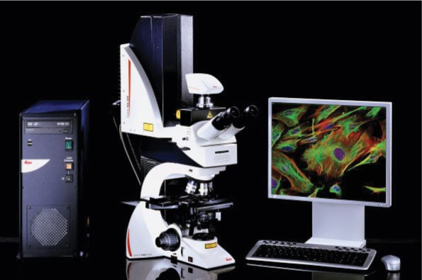
Leica Personal Confocal TCS SPE Upright Microscope
Olympus BX63 Upright Motorized Microscope Stereology System
High Performance Workstation with Cellsens® software
Workstation with Stereo Investigator® and Neurolucida® software
Management:
Chris Jurgens, Ph.D., Microscopy Core Director: Dr. Chris Jurgens received his Ph.D. from the University of North Dakota and completed postdoctoral training in Neurobiology at Virginia Commonwealth University in Richmond, VA. Dr. Jurgens brings extensive experience in DIC, fluorescence and other microscopy techniques.
Instrumentation and Facility Support:
The INBRE Microscopy Core is extensively equipped for the light microscopic examination of cells and tissues. Established and maintained by the ND INBRE program, this core is located in room W471 in the UND School of Medicine & Health Sciences building. A brief description of the equipment is detailed below.
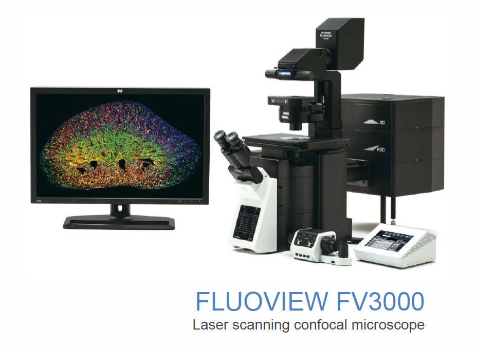
Olympus FV3000 Laser Scanning Confocal Microscope
The Olympus FLUOVIEW FV3000 series is an advanced confocal microscope designed to meet some of the most difficult challenges in modern science. The FV3000 is needed over and above the Leica TCS SPE due to the concentration of neuroscience research in ND. Much of the research ongoing in the neural system is focused on transcripts and proteins which occur in much less abundance that those normally studied in diseases such as cancer, heart disease, and diabetes. The FV3000 also allows the ND INBRE undergraduate training program to introduce students to the advantages of the Fluoview FV3000 compared to that of the personal confocal. The FLUOVIEW FV3000 is also equipped for live cell imaging.
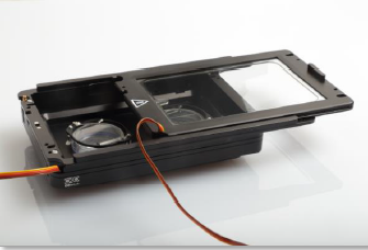
Live Cell Imaging
For live cell imaging, the Fluoview FV3000 is equipped with a removable a stage top incubation chamber consisting of an UNO Stage top controller for application with pre-mixed gases and an Olympus electrically heated chamber with sliding lid. The FV3000 also has silicone objectives (40x and 100x) and auto-focus for long-term time lapse imaging.
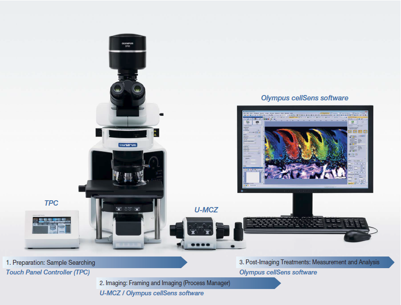
Olympus BX63 Upright Fluorescence Microscope
The Olympus BX63 Upright Fluorescence Microscopic System is a versatile high-end fluorescence imaging system. The BX63 is completely motorized and is uniquely focused via the nosepiece rather than stage, enabling the stage to be fixed making it more stable. The motorized stage is also a new feature and is driven by advanced, high-precision, ultrasonic Piezo technology, providing silent, smooth and extremely precise operation. The stage can even be positioned by hand enabling rapid gross sample alignment while highly accurate encoders continuously read-out the X and Y position. The excellent encoding enables the user to set precise coordinates and navigate directly to them at high speed.
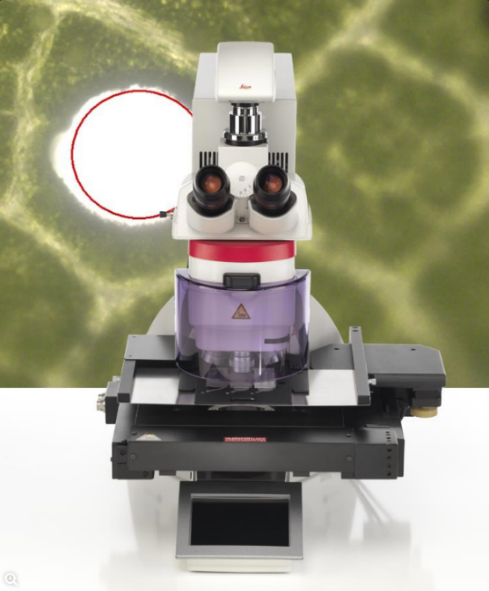
Leica LMD6 Laser Microdissection Microscope System
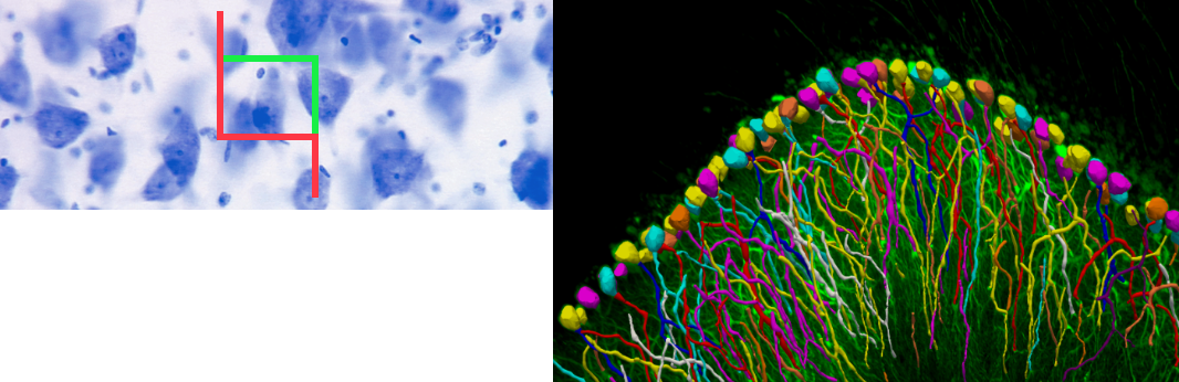
The Leica LMD 6 Laser Microdissection Microscope System is designed such that the laser is the element that moves and not the sample. Gravity is used for collection, dropping the sample contaminant free into the collection tube. The Leica LMD 6 is typically used in genomics (DNA), transcriptomics (mRNA, miRNA), proteomics, metabolomics, and even next generation sequencing (NGS). The system can be used for tissue, both frozen sections and sections from formalin-fixed, paraffin-embedded tissues, as well as cultured cells. The accompanying software package allows one to select, dissect, and visualize the dissectates, including photo documentation. The Leica LMD 6 system has been used successfully in ND INBRE workshops to introduce undergraduate and graduate researchers to the principles of histology and immunohistochemistry, with a final demonstration of the micro dissection of a student "hands-on" prepared slide of an ER+ breast cancer.
Leica Personal Confocal TCS SPE Microscope
The Leica Personal Confocal TCS SPE is an affordable entry level confocal for personal use or, very importantly, for undergraduate student training and unsupervised use by trained graduate students. The ND INBRE workshops have provided undergraduate students with a "hands on" demonstration of actin filament staining in cultured cells, visualization by confocal microscopy, and with a picture to own and show administrators, faculty, and other students their accomplishment.
Olympus BX63 Upright Microscope Stereology System
This stereology system consists of an Olympus BX63 Upright Motorized Microscope equipped with MBF Bioscience Stereo Investigator® and Neurolucida® software systems. These two software systems are powerful neuroanalytical tools. Stereo Investigator® offers a non-biased, automated approach to collecting data from tissue sections and is particularly valuable for multichannel labeled samples in z-series stacked images. This is achieved through precision control of the 3-axis stepping stage system that can mediate high throughput field-of-view scans. An important component of this software module is the ability to establish statistically significant collection boundaries that remove the component of investigator bias inherent in traditional neuroanatomical quantification. The sampling estimates and confidence values obtained are available through the software platform itself, or can be exported to other software programs. The Neurolucida® software system offers a flexible and interactive platform for conducting neuron tracing and reconstruction, neuroanatomical 3D mapping, and quantification of volumes and surfaces in the brain. This latter option is most important for developmental studies in which the 3D structure of the brain alters significantly over time. Neurolucida® and Stereo Investigator® also share the advantage of accepting data from a variety of sources including digital or analog images, confocal-derived z-series scans, and transmission and scanning electron micrographs. Each of the software systems offers a distinct quantitative or imaging advantage for stereological applications
Acknowledgement of ND INBRE Support:
Those using the Microscopy Core must agree to acknowledge ND INBRE support:
Research reported in this [publication, release] was supported by an Institutional Development Award (IDeA) from the National Institute of General Medical Sciences of the National Institutes of Health under grant number P20GM103442.
Technical and/or scheduling questions should be addressed to Chris Jurgens, christopher.jurgens@und.edu